Medical Microbiology Lecture: Bacterial Taxonomy, Cell Structure, and Gram Staining
AI summary of “Medical Microbiology Lecture: Bacterial Taxonomy, Cell Structure, and Gram Staining” by Microbiologia Marielsa Oficial, generated by Sumvid.
Title
Medical Microbiology Lecture: Bacterial Taxonomy, Cell Structure, and Gram Staining
One-Sentence Summary
This university lecture explains how bacteria are named and classified, contrasts prokaryotic and eukaryotic cells, describes the main bacterial structures and wall types, and walks through the Gram stain procedure and its clinical importance.
Worth Watching If
You are an introductory medical microbiology student who wants a visual, step-by-step overview of bacterial structure and the Gram stain. The delivery is rambling and the auto-generated transcript contains many errors, so readers who already know this material will find it basic.
Chapters
- [0:00] Introduction to taxonomy and why organisms are classified
- [1:03] Taxonomic ranks and binomial naming, using Bacillus subtilis as the example
- [4:14] Naming rules, Greek and Latin origins of names, and common nicknames for bacteria
- [5:49] Tree of life and the five kingdoms; bacteria as prokaryotes
- [7:24] Comparison table of prokaryotic and eukaryotic cells
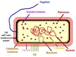
- [12:08] Internal and external bacterial structures: nucleoid, ribosomes, mesosomes, pili, flagella, membrane, wall, capsule
- [18:26] Peptidoglycan and the wall differences between Gram-positive and Gram-negative bacteria
- [24:44] Pili, capsules, and flagellar arrangements, with microscopy images
- [27:51] Endospores, with Bacillus and Clostridium as the key examples
- [33:05] Size comparison and bacterial shapes and arrangements
- [36:42] Gram-stain examples of cocci, bacilli, and other morphologies
- [42:03] Purpose and step-by-step procedure of the Gram stain
- [48:17] Lab setup, drying, and the need for oil immersion at 100x
Key Takeaways
- [0:33] Taxonomy exists to organize the enormous diversity of living things into nested groups: kingdom, phylum/division, class, order, family, genus, and species.
- [3:12] Bacterial scientific names use binomial nomenclature: the genus is capitalized, the species is lowercase, and the full name is italicized (or underlined or bolded).
- [7:24] Prokaryotic cells (bacteria and archaea) lack a nucleus and membrane-bound organelles, have circular DNA, and have 70S ribosomes. Eukaryotic cells have a nucleus and organelles, and their ribosomes are 80S (mitochondria and chloroplasts have 70S ribosomes).
- [8:25] Bacteria reproduce by binary fission, not mitosis, and can exchange genetic material through plasmids.
- [14:46] The capsule is a polysaccharide layer that protects against desiccation, phagocytosis, and antibody attack. The lecturer states that encapsulated strains are more virulent than non-encapsulated strains of the same species.
- [18:26] Peptidoglycan, made of N-acetylglucosamine and N-acetylmuramic acid, is the target of antibiotics such as beta-lactams and vancomycin. Because human cells lack peptidoglycan, these drugs are selectively toxic to bacteria.
- [20:01] Gram-positive bacteria have a thick peptidoglycan layer with teichoic acids. Gram-negative bacteria have a thin peptidoglycan layer between two membranes and an outer membrane containing lipopolysaccharide (LPS), which acts as an endotoxin.
- [28:24] Endospores are a survival structure, not a reproductive one. Bacillus and Clostridium are the two genera the lecture focuses on for spore formation.
- [36:42] Common morphologies include cocci, bacilli, and spirilla. Arrangements include diplococci (pairs), staphylococci (grape-like clusters), and streptococci (chains).
- [42:03] The Gram stain is the most important initial test in bacteriology because the result (Gram-positive or Gram-negative) guides the choice of antibiotic therapy.
- [45:09] Gram stain sequence: crystal violet (1 min), iodine mordant, alcohol-acetone decolorization, then safranin counterstain. Gram-positive cells retain the violet; Gram-negative cells lose it and turn pink/red. The order cannot be reversed.
Action Items
- Practice the Gram stain sequence in order, keeping each step at about one minute and rinsing with water between steps.
- Fix smears gently over a flame and let the slide dry completely before viewing.
- Use a drop of immersion oil when viewing at 100x, and make sure the slide is dry so the oil does not mix with water.
- When writing bacterial names, capitalize the genus, lowercase the species, and italicize the full name.
- Memorize the key differences between Gram-positive and Gram-negative walls, since they determine antibiotic selection.
Caveats
- The transcript is auto-generated and contains many errors in spelling and terminology (for example, "Neisseria" and some drug and organism names), so verify key terms against a textbook.
- The lecturer presents mesosomes as true membrane structures involved in respiration and DNA attachment. Many modern texts regard them as likely fixation artifacts.
- The statement that encapsulated bacteria are always more virulent is an oversimplification.
- Mycobacteria, which are mentioned, are usually identified by acid-fast staining rather than the Gram stain, and this distinction is not discussed.
- The description of the "fungus-like" actinomycetes responding to antibiotics is brief and may need context from a microbiology text.